A novel artificial intelligence model to improve detection of breast cancer metastasis has been developed by researchers at UT Southwestern Medical Center. The model could reduce the need for needle or surgical biopsies, UTSW said.
The noninvasive model uses standard magnetic resonance imaging (MRI), paired with machine learning AI, to detect axillary metastasis—the presence of cancer cells in the lymph nodes under the arms.
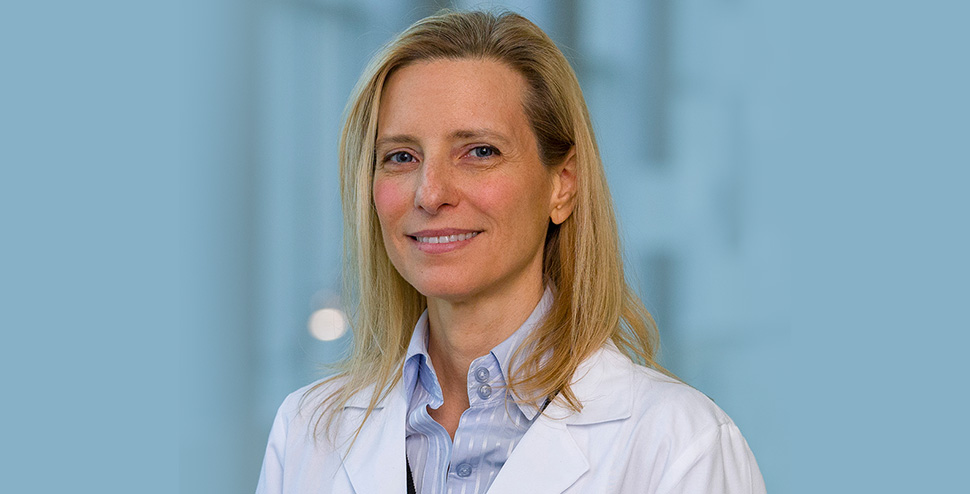
“Most breast cancer deaths are due to metastatic disease, and the first site is usually an axillary lymph node,” study leader Basak Dogan, M.D., professor of radiology, director of breast imaging research, and member of the Harold C. Simmons Comprehensive Cancer Center at UT Southwestern, said in a statement. “Determining nodal status is critical in guiding treatment decisions, but traditional imaging techniques alone do not have enough sensitivity to rule out axillary metastasis. That often requires patients to undergo invasive procedures that involve radioisotope and dye injection followed by surgery to remove and test whether the axillary nodes harbor cancer cells.”
Dogan is a Eugene P. Frenkel, M.D. Scholar in Clinical Medicine.
Training the AI model
The team’s research, published in Radiology: Imaging Cancer, showed that the AI model was significantly better at identifying patients with axillary metastasis than MRI or ultrasound, UTSW said. In clinical practice, the AI model would have helped avoid 51% of benign (noncancerous) or unnecessary surgical sentinel node biopsies while correctly detecting 95% of patients with axillary metastasis, UT Southwestern noted.
“That’s an important advancement because surgical biopsies have side effects and risks, despite having a low probability of a positive result confirming the presence of cancer cells,” Dogan said. “Improving our ability to rule out axillary metastasis during a routine MRI—using this model—can reduce that risk while enhancing clinical outcomes.”
UT Southwestern said the retrospective study used dynamic contrast-enhanced breast MRI exams from 350 newly diagnosed breast cancer patients at UTSW and the Moody Center for Breast Health at Parkland Health’s main campus in Dallas. All had known nodal status, UTSW said.
Those images, along with a range of clinical measures, were used to train the AI model to identify axillary metastasis using machine learning techniques.
Because the AI model is used along with standard imaging exams, it can also eliminate the stress and expense of additional testing for many patients.
“Patients with benign findings from traditional MRI exams or needle biopsies are often subjected to sentinel lymph node biopsy because those tests can miss a significant proportion of metastasis,” Dogan said. “Our research demonstrates that it’s possible to identify—with a high degree of accuracy—patients who are nonmetastatic, which benefits the patient and also allows the physician to tailor treatment.”
‘Great promise’ in the fight against cancers
UTSW said that the research builds on previous studies there related to breast cancer imaging and the development of predictive tools to detect metastasis.
“Our study is a testament to UT Southwestern’s commitment to impactful research that addresses real-world health care challenges,” Dogan said. “The development and validation of AI models for medical imaging holds great promise in helping us in the fight against breast and other cancers, and this new tool is a significant step forward.”
UTSW said the researchers continue to refine the image analysis process and are looking to include more varied data to validate their findings.
Researchers who contributed to the study are first author Dogan Polat, M.D., a second-year resident in radiology; Albert Montillo, Ph.D., assistant professor in the Lyda Hill Department of Bioinformatics and in Biomedical Engineering; Keith Hulsey, Ph.D., instructor of radiology; and Liqiang Wang, Ph.D., faculty associate, and Son Nguyen, Ph.D., postdoctoral researcher, in the Lyda Hill Department of Bioinformatics.
UTSW said the research was supported by the Simmons Cancer Center, the National Institutes of Health (NIH) National Institute of General Medical Sciences, NIH National Institute of Aging, NIH National Cancer Institute, the King Foundation, and the Lyda Hill Foundation.
![]()
Get on the list.
Dallas Innovates, every day.
Sign up to keep your eye on what’s new and next in Dallas-Fort Worth, every day.
R E A D N E X T
-
Included in the 12 Dallas-area funding awards is a $4 million Rising Star grant to bring Stefan Gloeggler, Ph.D., to UT Southwestern Medical Center from the Max Planck Institute of Multidisciplinary Sciences in Göttingen, Germany.
-
In a boost for bioentrepreneurs, the American Cancer Society’s BrightEdge has unveiled its new BrightEdge Entrepreneurs program. Aimed at accelerating the transition of cancer-focused innovations from research labs to the healthcare market, the program is set to begin on February 5, 2024. The 10-month initiative will support a cohort of 6 to 8 entrepreneurs, providing them with essential training, mentorship, and funding. The program targets scientific innovators who are ready to take their preliminary business ideas and develop them into viable companies dedicated to cancer diagnostics, therapeutics, medical devices, and digital health solutions. BrightEdge Entrepreneurs offers a rigorous curriculum addressing…
-
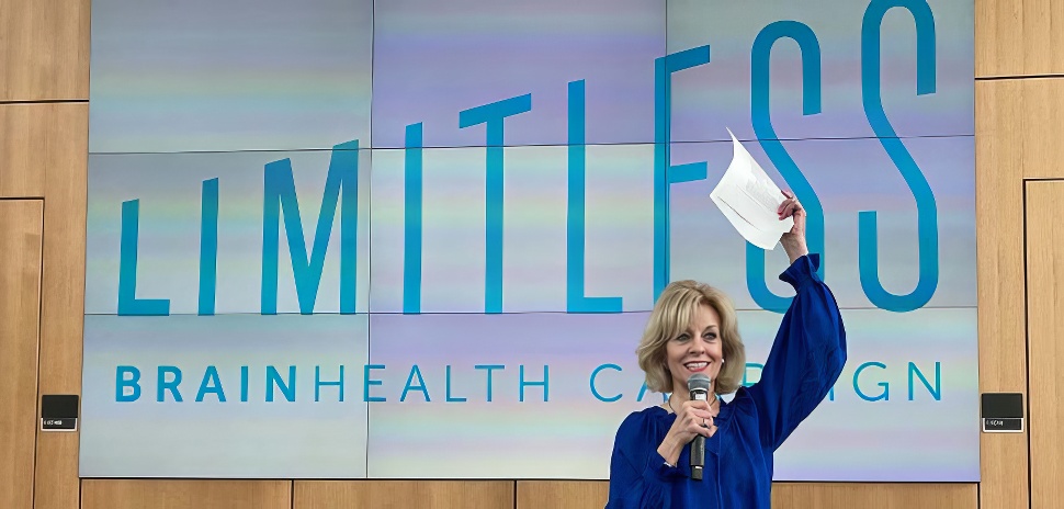
Now a quarter-century old, the Dallas-based cognitive neuroscience facility has grown from a "ragtag team" to a globally significant research institute dedicated uniquely to building better brain function. Here’s how it happened under chief director Sandra Bond Chapman, PhD.
-
Dallas-based Susan G. Komen—one of the world's leading nonprofits raising money for breast cancer research, awareness and community programs—has launched a cloud-based research platform specifically for breast cancer patients and survivors to share their personal health data with the organization. The data will be used to accelerate breast cancer research and help discover cures for breast cancer.
-
![Clockwise from left: Andrew Denton, Anisha Holla, Ralph Yongoueth, Ananya Sammidi, Asad Moulvi, Sahil Patel, and Rick Tett will pitch in UT Dallas' Big Idea Competition 2024 student and alumni track finals on April 17. [Photos via UT Dallas and the entrepreneurs; Graphics: DI Studio, istockphoto]](https://s24806.pcdn.co/wp-content/uploads/2024/04/UTD-Big-Ideas-Student-Alumni-Track-BIC2024.jpg)
The University of Texas at Dallas' largest startup pitch event, now in its 17th year, isn't sitting on its laurels. Here's what's new this year, and who made the finals. With dual events, a fireside chat, and judge-powered prize allocations, it's set to unfold on April 5 and 17.