RESEARCHERS SAY COFFEE MAY REDUCE CANCER RISKS
![]() I drink a lot of coffee, I admit it. I’m drinking some as I write. A new study from the Simmons Cancer Center at UT Southwestern Medical Center says that might be a good thing.
I drink a lot of coffee, I admit it. I’m drinking some as I write. A new study from the Simmons Cancer Center at UT Southwestern Medical Center says that might be a good thing.
Coffee consumption is steadily rising, UTSW said. When paired with prior studies showing that coffee consumption is associated with a lower risk of getting colon cancer, along with reduced risk of recurring tumors and death from colon cancer, one can take some solace in having that extra cup o’ joe in the morning, or in the afternoon, or the evening (decaf of course).
And speaking of decaf, the research shows that both regular and decaf work.
“We don’t quite know how coffee exerts its health benefit because there are many different compounds in coffee.”
Dr. Muhammad Beg
“We don’t quite know how coffee exerts its health benefit because there are many different compounds in coffee,” said Dr. Muhammad Beg, a GI cancer specialist at the Harold C. Simmons Comprehensive Cancer Center. “But researchers have shown that both caffeinated and decaf can be helpful.”
So, why don’t you grab a cup of your favorite coffee — arabica or robusta — and read more about the research here.
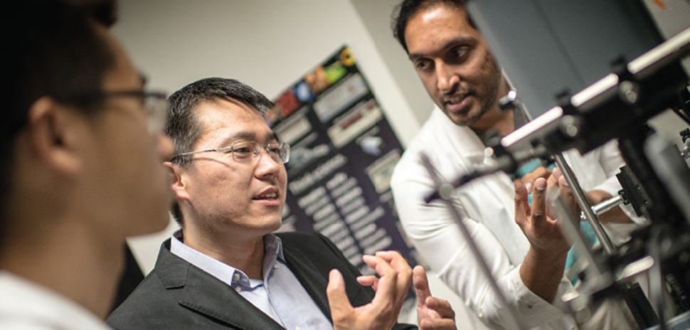
UT Arlington bioengineering professor Baohong Yuan, center, has spent years working on creating a better imaging system to detect cancer in deep tissues. [Photo courtesy of UT Arlington]
A $1.9M CPRIT GRANT IS FUNDING SCIENTIST’S WORK
Let’s be clear about it — one of the most-challenging pieces in the fight against deadly tumors is to capture clear pictures of them in deep tissues.
UT Arlington bioengineering professor Baohong Yuan has spent years working on creating a better imaging system to detect cancer that is tens of millimeters under the skin’s surface, according to UTA’s research website.
“The problem with current imaging is that what you get is not clear, but more like an out-of-focus photo.”
Baohong Yuan
Yuan’s work is being funded with two grants from the Cancer Prevention and Research Institute of Texas worth a total of $1.9 million.
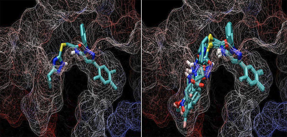
“The problem with current imaging is that what you get is not clear, but more like an out-of-focus photo,” Yuan told UTA Inquiry, the university’s research magazine. “For imaging of deeper tissues, you have to sacrifice resolution. This means if you want to see deep tissue, you cannot see too small. There’s a trade-off between the imaging depth and the resolution.”
Yuan is using ultrasound-mediated techniques that are combined with microparticles or nanoparticles that tumors attract to produce images of small, deep tumors.
His team has developed a non-invasive system that exposes the particles to ultrasound waves, making them temporarily fluorescent and, thus, detectable.
“We want to provide much clearer images of microvessels in deeply seated small tumors,” Yuan said. “With that information, physicians could better target the tumors for elimination.”
UT DALLAS RESEARCHER GETS MS SOCIETY GRANT
Bart Rypma of the Center for BrainHealth at the University of Texas at Dallas is one of 10 recipients who have been awarded funding from $433,800 in grants from the National Multiple Sclerosis Society.
Bart Rypma
Rypma’s study uses neuroimaging methods to identify mechanisms involved in MS-related cognitive dysfunction, according to UTD. The society said the 10 high-risk pilot projects are part of the yearlong Pilot Research Grant program which supports early stage research projects to quickly evaluate their effectiveness.
In 2016, Rypma was awarded more than $290,000 in grant funding from the society to investigate how changes in blood flow can affect cognition in people with MS.
3D PRINTING ‘IN-SITU’ RESEARCH GAINS MOMENTUM
In-situ 3D printing for bone regrowth is an emerging area of study by scientists looking at ways that 3D printers can aid in the healing process, according to a report in 3D Printing Industry.
“Our goal is to heal the defect or fracture site rapidly, as if nothing ever happened.”
Venu Varanasi
Dr. Venu Varanasi, assistant professor of biomedical sciences at Texas A&M University, recently presented at a session of the International & American Associations for Dental Research on bone regrowth utiliziing in-situ 3D printing — meaning printing in its original place.
“Our goal is to heal the defect or fracture site rapidly, as if nothing ever happened,” Varanasi said in the article.
He’s tested his approach on a cranial defect in rats where repairs were 3D printed and simulatenously cured under UV light in the upper part of the rat’s skull.
Varanasi’s research will be undertaken in the Bone-Muscle Group of the University of Texas at Arlington in collaboration with Texas A&M, 3 Printing Industry said.
Find out more here.
READ NEXT
Discovery: New UTSW Protocol Detects Bone Metastases, UTA Startup Gets Funding & Research Agreement
Discovery: Targeting Cancer Stem Cells, UTA Chemist Honored as Distinguished Scientist
Discovery: Aiding a Fragile Mussel, Trial Seeks Multiple Myeloma Patients, 3-D Universe
Discovery: Sway, TCU Partner on Sports Performance Research & UTA Gets $3.3M for Heart Study
![]()
Get on the list.
Sign up to keep your eye on what’s new and next in Dallas-Fort Worth, every day.
R E A D N E X T
-
After 10 years of working toward a more sustainable future worldwide, EarthX is doubling down on its move into television. The Dallas-based environmental organization is ramping up its EarthxTV streaming service with a new leadership team of TV veterans—with plans to eventually announce traditional and digital 24x7 linear distribution deals.
-
The Cancer Prevention and Research Institute of Texas (CPRIT) will convert previous grant awards to an equity investment. OncoNano uses pH as a biomarker to detect metastatic cancer. The technology "lights up" a tumor in imaging during real-time surgery.
-
It can take up to 15 years for a new drug to be available for the public to use. Through SMU's patent-pending ChemGen, researchers believe chemists may be able to "increase their productivity from working on as few as six problems a year to as many as 60."
-

In this weekly roundup of executive moves in North Texas, you'll also find news from Operation Kindness, Cooksey Communications, Munck Wilson Mandala, and StoneEagle F&I.
-
The investment was led by Advantech Capital, a PE fund based in China that focuses on TMT, pharmaceuticals, and healthcare. This combined with the support from the Cancer Prevention and Research Institute of Texas (CPRIT), which granted OncoNano $9.97 million last year, will support Phase 3 clinical trials for the biotech's technology that can diagnose and treat cancer with high specificity.