New 3-D software will produce holograms of the heart at Children’s Health Heart Centers in North Texas.
The technology will give family members a better understanding of their child’s condition, while helping physicians plan surgeries, D CEO Healthcare reports.
CANADIAN COMPANY DEVELOPED THE 3-D TECHNOLOGY
It’s especially helpful for difficult procedures because the images can be customized.
The 3-D heart mapping technology was developed by NGRAIN, a Canadian firm.
First, the heart is scanned through an MRI or similar procedure. Then, software converts those images into 3-D.
“We can be sure that the position is perfect for the intervention we want to do.”
Dr. Tarique Hussain
“It’s when we have a difficult procedure and the surgical decision-making is difficult, this kind of 3-D planning really helps,” said Dr. Tarique Hussain, a pediatric cardiologist at Children’s Health. “We can be sure that the position is perfect for the intervention we want to do.”
It also means less radiation is needed during the procedure.
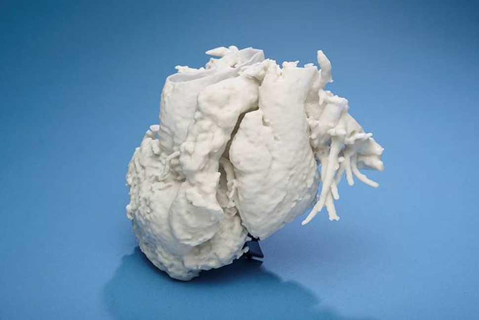
That data can even be printed using a 3-D printer so there will be a tangible model. Children’s Health has its own 3-D printer and can also access ones at UT Southwestern, which is also at the cutting edge of 3-D holograms, and at the University of Texas at Dallas.
Drs. Gerald Greil and Animesh Tandon at Children’s Health’s Heart Center have pioneered the research into the surgical applications of 3-D technology.
In one case, there was a heart defect, and a stent needed to be placed in the child’s heart. The 3-D printed heart allowed them to find a minimally invasive way of doing it.
“It’s a novel procedure and the 3-D printing gave us the confidence that the procedure would work.”
Dr. Tarique Hussain
“For a lot of these cases, we would be able to deal with the defect without doing surgery,” Hussain said. “It’s a novel procedure and the 3-D printing gave us the confidence that the procedure would work.”
The use of 3-D technology will only increase throughout the medical field.
“It will change the way doctors discuss the cases–moving from 2-D imaging to 3-D imaging and eventually to 3-D holograms of the heart,” Hussain said.
The models can be used as teaching tools for fellow physicians and medical students.
https://www.facebook.com/Childrens/videos/10154493960728137/
Related reading:
Bioinks at Heart of 3-D Printing Human Tissue, Organs
Delivering what’s new and next in Dallas-Fort Worth innovation, every day. Get the Dallas Innovates e-newsletter.

